Comprehensive Eye Exam
A comprehensive eye exam is a thorough evaluation of your eyes and vision, aiming to detect any eye problems at their earliest stages when they’re most treatable.
What to Expect During a Comprehensive Eye Exam:
A specially trained ophthalmic technician will work with you to complete many components of the exam before your time with the doctor. They begin with reviewing and confirming your medical history, documenting current medication and eyeglass prescriptions, as well as asking about symptoms or eye problems you are experiencing. They will be performing preliminary tests to optimally assess your eye health for the doctor’s evaluation.
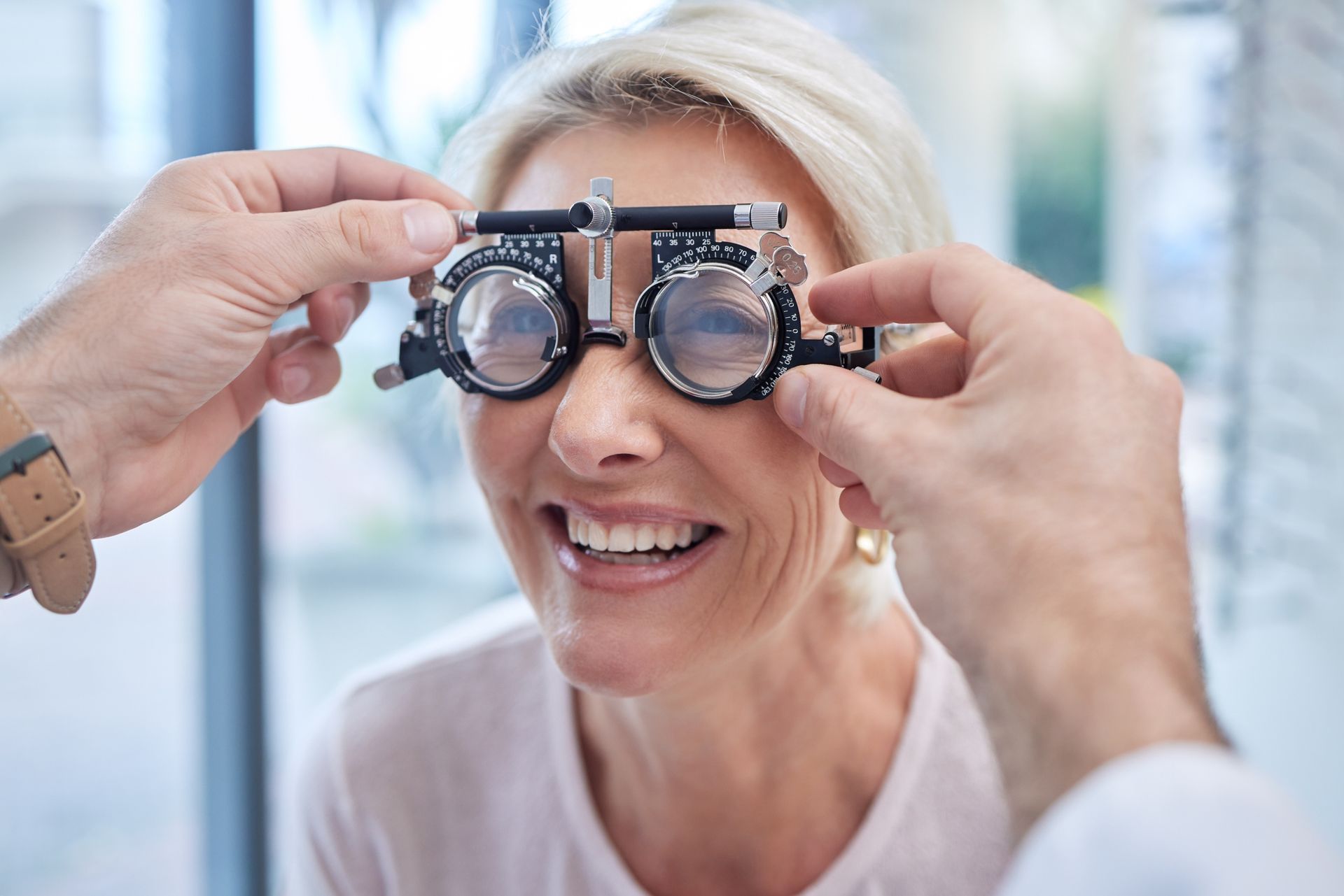
The Refraction Test
The purpose of conducting a refracting test is to determine your best possible visual acuity. The information obtained during a refraction allows us to provide you with a new prescription, if necessary. A specially trained ophthalmic technician will show you a series of lens choices through the phoropter and ask you to compare to them by looking at a chart usually placed across the room. For each pair of lenses, you will be asked, “Which looks clearer, option one or option two?” Based on your responses, the eye doctor fine-tunes the lens power until reaching the best possible clarity for your vision. The process is repeated for each eye individually.
The Tonemetry Test
The tonometry test is a preventive measure that can play a crucial role in protecting your vision. This test helps your doctor detect glaucoma early on by measuring the pressure inside your eyes. We do this by a quick, straightforward, painless process.
Comprehensive Examination
The eye health examination is comprised of many components; each part of the eye will be examined detail, using specialized equipment.
Anterior Examination
When going through an anterior examination, or the front part of your eye, special instruments, such as slit lamps and ophthalmoscopes, are used to illuminate and magnify different parts of your eye. Specialized equipment are used to check the structure of the eyelids, cornea, iris and lens, for any signs of disease or conditions that might affect your vision or eye health.
Posterior Examination
To examine the posterior, or back part of the eye, a more in-depth look is necessary. This area includes the retina and optic nerve. There are two ways to views this area, dilation and photographic imaging.
Dilation
Your doctor may use eye drops to dilate (widen) your pupils. This temporary effect enlarges your pupils, allowing more light to enter and providing a clearer view of the back of the eye, including the retina, optic nerve and blood vessels.
It is a temporary effect and many people bring sunglasses to their appointment to wear afterward, reducing discomfort from bright lights.
Alternatively, a special camera can take detailed images of your retina without need for dilation. This method provides a clear picture of your eye’s health and is specifically useful for monitoring changes over time.